 Subclavian artery - Wikipedia
Subclavian artery This article needs additional appointments for . Please help. Unsolicited material can be challenged and disposed of. Find sources: – · · · · (January 2020) () you need additional quotations for ··· Subclavian artsSchematic of the proximal aorta and its branches. The left subclavian artery is the fifth branch of the aorta and the third branch of the aorta arch. The right subclavian artery emerges from the branches and their branches. (The right subclavian is at the top left, and the left subclavian is at the top right.) DetailsSource (left) (right)Branches (mainly)Identifiersarteria subclavian[]In , the subclavian arteries pair more of the upper part, below the . They receive blood from the . The left subclavian artery supplies blood to the left arm and the right subclavian artery supplies blood to the right arm, with some branches supplying the head and chest. On the left side of the body, the subclavian leaves directly from the , while on the right side it emerges from the relatively short when it forks in the subclavian and the right . subclavian arteries The usual branches of the subclavian on both sides of the body are , , , , el and , which can branch of the transverse cervical artery, which is a branch of the thyrocervical trunk. The subclavian becomes the side edge of the first. ContentEstruture[]From its origin, the subclavian artery travels laterally, passing between previous and medium, with the previous staircase () on its previous side and the medium scale () on its back. This is in contrast to the , which travels before the previous scalpel. As the subclavian artery crosses the lateral border of the , it becomes the . On the right side the subclavian artery emerges from the brachiocephalic artery (innominated) behind the right sternoclavicular joint; on the left side it springs from the aorta arch. The two vessels, therefore, in the first part of their course, differ in length, direction and relationship with the neighboring structures. The left subclavian artery is about 6 long in , while the right subclavian artery is about 9 cm long. Both have a width of 9-12. Parties[]To facilitate the description, each subclavian artery is divided into three parts: The first portions of the two vessels require separate descriptions; the second and third parts of the two arteries are practically equal. Part One[]The first part of the right subclavian artery emerges from the brachyocphalic trunk, behind the upper part of the right sternoclavicular joint. It passes up and upwards the middle margin of the . It rises a bit above the middle part of the . It is covered, in front, by the integument, superficial fascia, the , deep , the clavicular origin of the , , and , and another layer of . It is crossed by the and , by the cardiac and vague branches, and by the subclavian loop that forms a ring around the vessel. He is headed laterally in front of the artery, but separated from it by the sternohyoide muscle and the sternotiroid muscle. Below and behind the artery is the , which separates it from the apex of the . Behind the artery is the sympathetic trunk, the first and the first (T1). Winds around the lower and back of the ship. The first part of the left subclavian artery emerges from the , behind the , and at the level of the . It rises in the root of the neck, and then arches toward the side toward the medial border of the previous step muscle. It is in relation, in front, with the vague nerve, the heart nerves and the frenic nerves, which are parallel to it, the common carotid artery left, the internal left and vertebral yugular veins, and the beginning of the unnamed vein left. It is covered by the sternothyroid muscle, the steroid muscle and the sternocleidomastoid muscle. Behind it, it is in relation to the esophagus, chest duct, left recidivating larynx nerve, lower cervical node of the sympathetic trunk, and the colli longus muscle; higher, however, the esophagus and the chest duct are on their right side; the latter ultimately arching on the container to join the binding angle between the inner and subclavian veins. Medial to it are the esophagus, trachea, chest duct and the left recurring larynx nerve. Later they are the left pleura and the lung. Part Two[]The second part of the subclavian artery is behind the former scalnus muscle and in front of the . It is very short, and forms the highest part of the arch described by the container. In front, it is covered by the skin, the surface fascia, the platysma muscle, the deep cervical fascia, the sternocleidomastoid muscle, and the previous scaly muscle. On the right side of the neck, it is separated from the second part of the artery by the anterior scalnus muscle, while on the left side crosses the first part of the artery near the mid edge of the muscle. Behind the artery are pleura and medius escalnus muscle. Above the artery is the . Below the artery is the pleura. The subclavian vein is below and prior to the artery, separated from it by the previous muscle steps. Part III[]The third part of the subclavian artery runs down and downwards from the lateral margin of the anterior scayola muscle to the outer edge of the first rib, where it becomes the axillary artery. This is the most shallow part of the vessel, and is contained in the subclavian triangle. It is covered, in front, by skin, superficial fascia, platysma muscle, supraclavicular nerves and deep cervical fascia. The outer jugular vein crosses its medial part and receives the cross-sectional, cervical and previous yugular veins, which often form a plexo in front of the artery. Behind the veins, the Subclavian nerve goes down in front of the artery. The terminal part of the artery is behind the clavicle and the Subclavian and is crossed by the transverse scapular vessels. The subclavian vein is ahead and at a slightly lower level than the artery. Behind, it is found in the lower trunk of the brachial plexo, which intervenes between it and the medius squamous muscle. Above and on the side are the upper trunks of the brachial plexus and the omohioid muscle. Then rest on the upper surface of the first rib. Branches[] The subclavian arteries give five main arteries each: , , , , , , and . Part Branches Course Part one From its origin to the medial border He runs cranially on the , joins the vertebral artery on the counterlateral side, forming the and joins the . It runs well behind the ribs, giving, piercing the glasses to the and ending in the and the . Very short. It is divided into , and (also called cervicodorsal trunk). Part two lying behind previous steps It divides into and. Part three Between the lateral frontier of previous scales and the outer border of the From the second or third part. Go back to supply and remboid. Part One From its origin to the median border Part TwoLying behind scalenus previous Part III Between the lateral frontier of previous scales and the outer border of the Development[], the left subclavio simply arises from the 7th left, while the right subclavio emerges, proximal to the distal: Essentially, the fourth aortic arch and dorsal aorta form the aortic arch of the left, but since the right dorsal returns distal to the 7th intersecmental right artery, to the left form the proximal part of the subclavian artery. Since the left subclavian is then a tributary of the common left carotid, you can think that it arises from the brachiophalic trunk. Variation[] Subclavian arteries vary in their origin, course and height to which they rise in the neck. The origin of the right subclavian of the unnamed takes place, in some cases, above the sternoclavicular joint, and occasionally, but less frequently, below that joint. The artery may arise as a trunk separated from the aorta bow, and in such cases it may be the first, second, third, or even the last branch derived from that vessel; in most, however, it is the first or last, rarely the second or third. When it is the first branch, it occupies the ordinary position of the unnamed artery; when the second or third, it gains its usual position passing behind the right carotid; and when the last branch arises from the left extremity of the arch, and passes obliquely to the right side, usually behind the trachea, esophagus, and the right carotid, sometimes between the esophagus and the first. In very rare cases, this glass comes from the chest aorta, as low as the fourth thoracic vertebrae. Occasionally, pierce the previous Scalenus; more rarely passes in front of that muscle. Sometimes the subclavian vein passes with the artery behind the previous ladder. The artery can ascend up to 4 cm above the collarbone, or any intermediate point between it and the upper edge of the bone, the right subclavian usually ascending higher than the left. The left subclavian occasionally joins its origins with the left common carotid artery, forming a left brachiophalic trunk. The left subclavian artery is more deeply placed than the right on the first part of its course, and as a rule it does not reach such a high level in the neck. The later border of the Sternocleidomastoideus corresponds quite close to the lateral border of the previous Scalenus, so that the third part of the artery, the most accessible part for the operation, is immediately side to the later border of the Sternocleidomastoideus. Some authors describe the subclavian artery as a result of the seventh intersecmental artery. Function[] Subclavian arteries carry most of the blood that supplies the arms. It also supplies blood to the neck and brain. Clinical Significance[]The compression of the subclavian artery may cause (TOS). Subclavian arteries can be vulnerable to . occurs when there is occlusion or stenosis of the subclavian artery at a point before the branching of the . This can cause blood to flow through the vertebral artery into the distal subclavian artery, allowed by reduced pressure. Subclavian arteries are relatively shallow, and can be seen using. Additional images[]Side of neck, showing chief surface markings. MRI scan Right subclavian artery Brachial duplex and subclavian artery See also[]References[ ]This article incorporates text in the 20th edition of (1918) abcdefghijklmnabcabcdefabcdefghijklmnopabab External links[] Wikimedia Commons has media related to . ########################################################################################################################################################################################################################################################## 1st part / 2nd part / 3rd part / / ocular group: 1st part / 2nd part / 3rd part / / ocular group: 1st part / 2nd part / 3rd part / 1st part / 2nd part / 3rd part / / ocular group: ocular group: of the lungs Heart Sections Sections Navigation menu Personal tools Named spaces Variants Views More Search Navigation Contributed Tools Printing/exporting Other projects Languages
Server Error Please try later. Accessibility links Search results Subclavian arteclava - Wikipedia Subclavian arteclava: Anatomy, branches and clinical notes... subclavian artery: Anatomy, branches and nenemonic tention Kenhub Subclavian artery – How subclavian artery - YouTubeSubclavian branches - Structure Detail - anatomyEXPERTSubclavia Arteria Subclavia
 VIT(1st part) C (2nd part) D(3rd part, Dorsoscapular artery) [ Note:… | Subclavian artery, Arteries anatomy, Arteries")
Branches of parts of Subclavian artery ... (*) VIT(1st part) C (2nd part) D(3rd part, Dorsoscapular artery) [ Note:… | Subclavian artery, Arteries anatomy, Arteries
Subclavian Arteries | The Lecturio Medical Online Library:background_color(FFFFFF):format(jpeg)/images/article/en/the-subclavian-artery-and-its-branches/59X0N5FNUENPET04ADTDaw_kFnFtOY3FbNzz36Nsbdg_Subclavian_artery-2.png "Subclavian artery: Anatomy, branches and clinical notes | Kenhub")
Subclavian artery: Anatomy, branches and clinical notes | Kenhub
The Anatomy of the Laboratory Mouse
Subclavian Artery Stenosis | Thoracic Key
Anatomy - Subclavian artery branches - YouTube
Easy Notes On 【Subclavian Arteries】Learn in Just 3 Minutes! – Earth's Lab
Instant Anatomy - Upper Limb - Vessels - Arteries - Subclavian artery
Subclavian Artery Branches / Mnemonic Series #14 - YouTube
subclavian artery branch - Google 搜尋 | Carotid artery, Vertebral artery, Arteries
Subclavian Artery - The Definitive Guide | Biology Dictionary
Acute Subclavian Artery Thrombosis - Hand - Orthobullets
Subclavian artery and it's branches
Easy Notes On 【Subclavian Arteries】Learn in Just 3 Minutes! – Earth's Lab
Subclavian Artery , Origin, Branches and Relations , Anatomy QA
The subclavian artery and its branches have been demonstrated. (SA:... | Download Scientific Diagram:background_color(FFFFFF):format(jpeg)/images/library/12428/Brachial_plexus_roots_and_neck_viscera.png "Subclavian artery: Anatomy, branches and clinical notes | Kenhub")
Subclavian artery: Anatomy, branches and clinical notes | Kenhub![Figure, Aorta, Right Common Carotid Artery,...] - StatPearls - NCBI Bookshelf](https://www.ncbi.nlm.nih.gov/books/NBK499911/bin/aorticArch.jpg "Figure, Aorta, Right Common Carotid Artery,...] - StatPearls - NCBI Bookshelf")
Figure, Aorta, Right Common Carotid Artery,...] - StatPearls - NCBI Bookshelf
Thorax:Arterial structure:Subclavian arteries | RANZCRPart1 Wiki | Fandom![Презентация на тему:]()
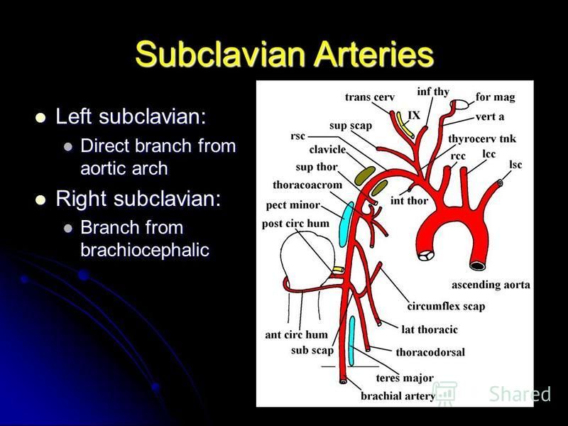
Презентация на тему: "VASCULAR SUPPLY TO UPPER EXTREMITY ARTERIES. Subclavian Arteries Left subclavian: Left subclavian: Direct branch from aortic arch Direct branch from aortic.". Скачать бесплатно и без регистрации.
Subclavian artery | Radiology Reference Article | Radiopaedia.org
Subclavian Artery , Origin, Branches and Relations , Anatomy QA
What are the branches of the Subclavian artery? Mnemonic - Anatomy - YouTube
Acute Subclavian Artery Thrombosis - Hand - Orthobullets
New findings of branching variations in subclavian arteries and supra-aortic arteries in Felis catus | SpringerLink - Atlas of Surgical Techniques in Trauma")
Subclavian Vessels (Chapter 9) - Atlas of Surgical Techniques in Trauma
Aberrant right subclavian artery | Radiology Reference Article | Radiopaedia.org
Subclavian artery - Wikipedia
Axillary artery branches... 【 Axillary artery is divided into 3 parts by - Pectoralis minor while Subclavian artery… | Arteries anatomy, Subclavian artery, Arteries
Anatomy of the Cerebral Structure | Radiology Key
Medicowesome: Branches of subclavian artery mnemonic
Subclavian Arteries | The Lecturio Medical Online Library
Right Subclavian Artery - an overview | ScienceDirect Topics
The subclavian arteries | Musculoskeletal Key
The branches of the left subclavian artery in the rabbit 1-left... | Download Scientific Diagram
Easy Notes On 【Subclavian Arteries】Learn in Just 3 Minutes! – Earth's Lab
Subclavian artery parts and branches Diagram | Quizlet
Branches of Subclavian artery Mnemonics
branches of subclavian artery | Arteries anatomy, Internal carotid artery, Subclavian artery
Pharyngeal branches subclavian


Posting Komentar untuk "branch of subclavian artery"